

Data and Evidence
Reduction in Seroma and Other Complications with a Novel Internal Negative Pressure System in Breast Reconstruction
Dr. Robert Paul, MD
Plastic and Reconstructive Surgery – Global Open, September 2023 – Volume 11, Issue 9 - p e5261
Click here to download a copy of the paper
This retrospective study using IC Surgical’s Interi™ System in prepectoral breast reconstruction demonstrated a significant reduction in seroma and other complications compared to standard drains.
Statistically significant differences were reported in four areas:
| Interi n=170 breasts |
Standard drains n=166 breasts |
P |
|
|---|---|---|---|
| Seroma | 7 (4.1%) | 38 (22.9%) | <0.00001 |
| Skin/flap revision | 18 (10.6%) | 36 (21.7%) | 0.006 |
| Any complication | 40 (23.5%) | 73 (44.0%) | 0.0001 |
| Duration of therapy | 16.5 days ± 3.5 | 19.6 days ± 6.2 | <0.0001 |
Dr. Paul concludes the observed reductions in mastectomy skin flap revisions and seroma are indicitive of improved internal wound healing. Interi is effective across a broad spectrum of patients, including high risk patients.
Abstract
Seroma, along with other complications, occurs as a result of poor wound healing following breast reconstructive surgery. Initial experience in a small cohort undergoing prepectoral breast reconstruction showed a clinical and statically significant reduction in seroma and any complication versus standard drains. The purpose of this study is to report on the safety and effectiveness of Interi compared with standard drains, in a larger patient population followed up over a longer period than the initial study.
Methods: Data on demographics, mastectomy and reconstructive variables, postoperative complications, and manifold/drain duration were retrieved from patient records and compared between the two groups.
Results: Interi was used in 100 patients (170 breasts) and standard drains in 100 patients (166 breasts). Groups were well matched in all demographics, reconstructive, and mastectomy variables. Interi was removed significantly earlier than drains (16.5 vs 19.6 days; P=<0.0001) and was associated with a significantly lower incidence of seroma (4.1% versus 22.9%, P<0.00001), flap revision (10.6% versus 21.7%, P=0.006), and any complication (23.5% versus 44.0%, P=0.0001).
Conclusion: Interi effectively reduced dead space and evacuated fluid from internal tissue planes, thereby decreasing seroma and other complications after prepectoral breast reconstruction. As a viable alternative to standard drains, it could significantly improve patient outcomes.
The Internal Negative-Pressure Wound Control System: A Paradigm Shift for Promoting Deep Space Healing in Complex Surgically Created Wounds
Dr. Kenneth C. Shestak, MD
Aesthetic Surgery Journal, Volume 41, Issue 11, November 2021, Pages NP1543–NP1549
Click here to access the paper from publisher’s website
Dr. Shestak’s paper establishes the historical problem of poor post-surgical internal healing and introduces IC Surgical's Interi® System as a transformative new technology, designed to address this major unmet need in surgery. This paper includes results from a 24-patient pilot clinical study in abdominoplasty using Interi technology.
Abstract
This article introduces a new technology to minimize seroma and promote more predictable healing in surgically created deep space wounds. Its novel design internalizes the delivery of a continuously generated high negative pressure (–125 mmHg) throughout the surgically created space by means of a multibranched Manifold. In a small prospective cohort case study of 24 patients undergoing full abdominoplasty, all patients underwent placement of this device, which was removed 7 days postoperatively. Results at 30 days revealed no evidence of wound-healing problems, no clinical seroma, and no device malfunction. The internalization of a constant negative-pressure wound therapy provided by this system has the potential to significantly reduce clinical seroma, and to produce more consistent apposition of interfaces in deep tissue spaces in complex wounds seen in plastic surgery and other surgical disciplines.
Pilot Clinical Study
Prospective Pilot Study to Evaluate Initial Clinical Safety and Functionality of the Internal Tissue Closure System in Patients Receiving Anterior Full Abdominoplasty
IC Surgical completed a successful prospective pilot clinical study demonstrating the absence of seroma, infection, or other wound complications using the IC Surgical technology. The clinical study enrolled 24 patients undergoing anterior full abdominoplasty (“tummy tuck”) at three sites in the U.S. All patients successfully completed the study including 30-day follow-ups.
Study Objectives
- Primary: To evaluate clinical safety of the system
- Secondary: To evaluate functionality of the system
Investigators
Brad Bengtson, MD, FACS, Grand Rapids, MI
Max Lehfeldt, MD, FACS, Pasadena, CA
Jan Turkle, MD, FACS, Indianapolis, IN
Study Design
Following surgeon standard procedure for abdominoplasty incision and tissue dissection, the peel apart section of the Manifold was positioned in the surgically created open tissue plane with full separation and placement of the 4 branches of the peel apart section of the Manifold (Picture 1). The distal end of the Manifold tubing was tunneled through soft tissue to an exit point. The portable suction pressure device was attached and activated in the OR.
Patients were discharged with the suction device connected and instructed to monitor fluid levels and exchange the portable suction device when full. The Manifold remained in place connected to the suction device for no less than seven days. At day 7, If fluid removal was <20 mL in preceding 24 hours, the Manifold was removed by the surgeon through the exit site. Surgical and exit site assessment and pain assessment were completed during 4 post-operative visits. Patients were followed for 30 days.
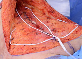
Picture 1 - Manifold branches placed in the abdominoplasty open tissue plane
Results
The pilot clinical study was effective at demonstrating its primary safety objective, with no safety events reported. No adverse events were reported including no skin edge necrosis, no wound dehiscence and no wound infections. Importantly, there were no clinical seromas reported on physical exam in any patient.
Further, the pilot clinical study also demonstrated effective post-operative fluid removal, closing down internal tissue planes, and was well-tolerated by patients with minimal to no pain upon removal. Subjectively, nursing staff and investigators noted relatively less visible edema of overlying skin flaps than is typically observed in abdominoplasty patients.
| Subject information | |
|---|---|
| Number of subjects enrolled | n=24; 100% female |
| Average age | 40.6 years (Range: 25 – 63) |
| Average BMI | 25.1 (Range: 20 – 29) |
| Average volume of fluid removed | 551 mL (Range 240 – 2205 mL) |
| Intra-operative data | |
|---|---|
| Average flap size removed | 764 cm2 (Range: 288 – 1350 cm2) |
| Average flap dimensions | 25.9 cm x 28.8 cm (Range: 18 cm x 16 cm - 30 cm x 45 cm) |
| Days of removal assessments | |
|---|---|
| Removal at POD 7 | 100% of subjects |
| “The system was effective in management of post‑operative fluid removal” | Subjects: 100% Strongly Agree (n=24) Surgeons: 96% Strongly Agree, 4% Agree (n=24) |
| “The system functioned acceptably during duration of therapy” | Subjects: 100% Strongly Agree (n=24) Surgeons: 100% Strongly Agree (n=24) |
| Subject Assessment of Pain Upon Removal | Average assessment score: < 1 88% scored 0 or 1 “None” or “Little Bit”, no score above 2 based on a visual analog scale 0=”No Hurt” to 5=”Worst Hurt” |
| Ultrasound images |
|---|
High resolution ultrasound was performed in a subset of patients, with no fluid collection detectable in these patients at any postop visit, including post-op Day 30. Representative ultrasound images from a single patient on Day 7 and Day 30 (Picture 2 & 3) show internal tissue planes without seroma or fluid collection.
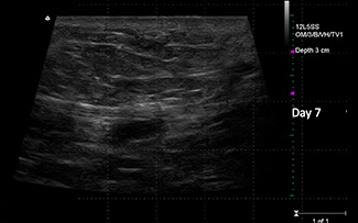
Picture 2 - Representative ultrasound image from Post-op Day 7
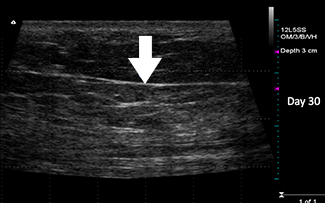
Picture 3 - Representative ultrasound image from Post-op Day 30
Pre-clinical studies
Development of the Interi System involved an iterative series of pre-clinical studies to demonstrate effectiveness of the platform concept and inform design of the proprietary system.
Design of studies
Key areas of investigation in these studies involved the creation of surgical pockets in a porcine model. A series of 12 animal studies were implemented to demonstrate system effectiveness, including 77 total subjects.
The following aspects of the technology were tested in the animal model:
Healing response to various materials
Manifold design to achieve fluid removal specifications
Manifold design to achieve tissue closure
Presence and duration of tissue adherence
Manifold removal force
Appropriate suction pressure ranges
Results
| Key findings related to Manifold |
|---|
| Demonstrated biocompatibility |
| Confirmed material strength |
| No infiltration of tissue |
| Removal force same as standard of care |
| Key findings related to overall system |
|---|
| Showed effectiveness of the therapy |
| Achieved fluid removal expectations |
| Gross observations |
|---|
Two surgical tissue planes were created in each animal, to compare location treated with standard of care and location treated with IC Surgical technology. Explants were performed at 3 months, with visual wound assessment performed on explanted tissue. Representative cross-sections show explanted tissue treated with standard of care (Picture 4) and treated with IC Surgical technology (Picture 5), with tissue apposition observed in the IC Surgical test group.
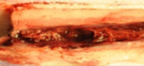
Picture 4 - Visual assessment of explanted tissue treated with standard of care demonstrates lack of tissue apposition with gaps for fluid accumulation and weakened repair.
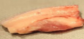
Picture 5 - Visual assessment of explanted tissue treated with IC Surgical technology demonstrates closed tissue planes and evidence of healing.
Conclusion from Clinical and Pre-clinical Studies
Early evidence supporting the technology performance in clinical and pre-clinical studies supports our intended goal of providing the surgeon with internal wound control to optimize tissue restoration.